Caterina Porcino, Marilena Briglia, Marialuisa Aragona, Kamel Mhalhel, Rosaria Laurà, Maria Levanti, Francesco Abbate, Giuseppe Montalbano, Germana Germanà, Eugenia Rita Lauriano, Alessandro Meduri, Josè Antonio Vega, Antonino Germanà, and Maria Cristina Guerrera
Published: 6 January 2023 | Molecular Neurobiology

Abstract
The incidence rates of light-induced retinopathies have increased significantly in the last decades because of continuous exposure to light from different electronic devices. Recent studies showed that exposure to blue light had been related to the pathogenesis of light-induced retinopathies. However, the pathophysiological mechanisms underlying changes induced by light exposure are not fully known yet. In the present study, the effects of exposure to light at different wavelengths with emission peaks in the blue light range (400–500 nm) on the localization of Calretinin-N18 (CaR-N18) and Calbindin-D28K (CaB-D28K) in adult zebrafish retina are studied using double immunofluorescence with confocal laser microscopy.
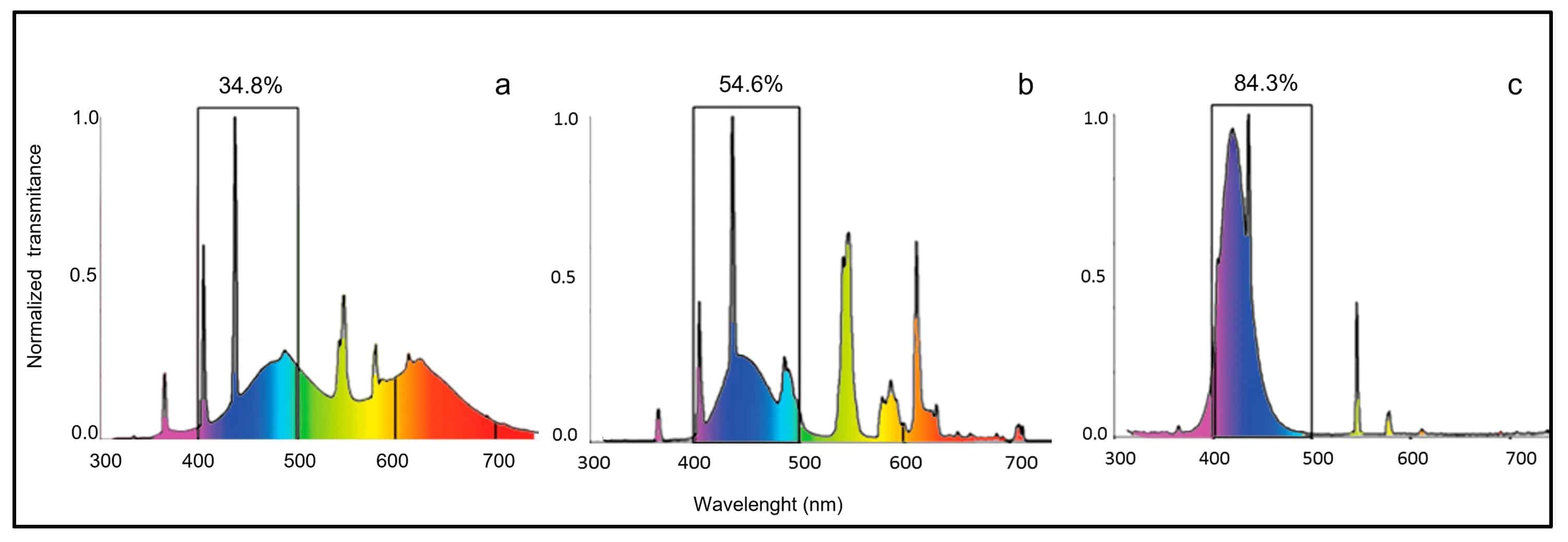
Figure 5. Spectral distribution of the illumination sources. (a) white light, (b) white–blue light, (c) blue light. The percentage of blue light emission for each experiment is indicated.
CaB-D28K and CaR-N18 are two homologous cytosolic calcium-binding proteins (CaBPs) implicated in essential process regulation in central and peripheral nervous systems. CaB-D28K and CaR-N18 distributions are investigated to elucidate their potential role in maintaining retinal homeostasis under distinct light conditions and darkness. The results showed that light influences CaB-D28K and CaR-N18 distribution in the retina of adult zebrafish, suggesting that these CaBPs could be involved in the pathophysiology of retinal damage induced by the short-wavelength visible light spectrum.
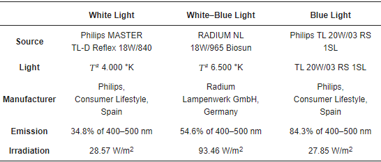
… Table 3. The spectral composition of the light source was determined using the optic fiber spectrophotometer Spectrawiz EPP2000 (StellarNet, Keystone, FL, USA) …


