
What is Optical Coherence Tomography (OCT)?
Optical Coherence Tomography (OCT) is a non-invasive imaging technique that captures high-resolution, cross-sectional images of biological tissues. It utilizes light waves to take micrometer-resolution, two- and three-dimensional images, making it a powerful tool for detailed tissue examination. OCT is widely explored in research and medical diagnostics for its potential to revolutionize the detection and monitoring of diseases such as glaucoma, macular degeneration, and cardiovascular conditions.
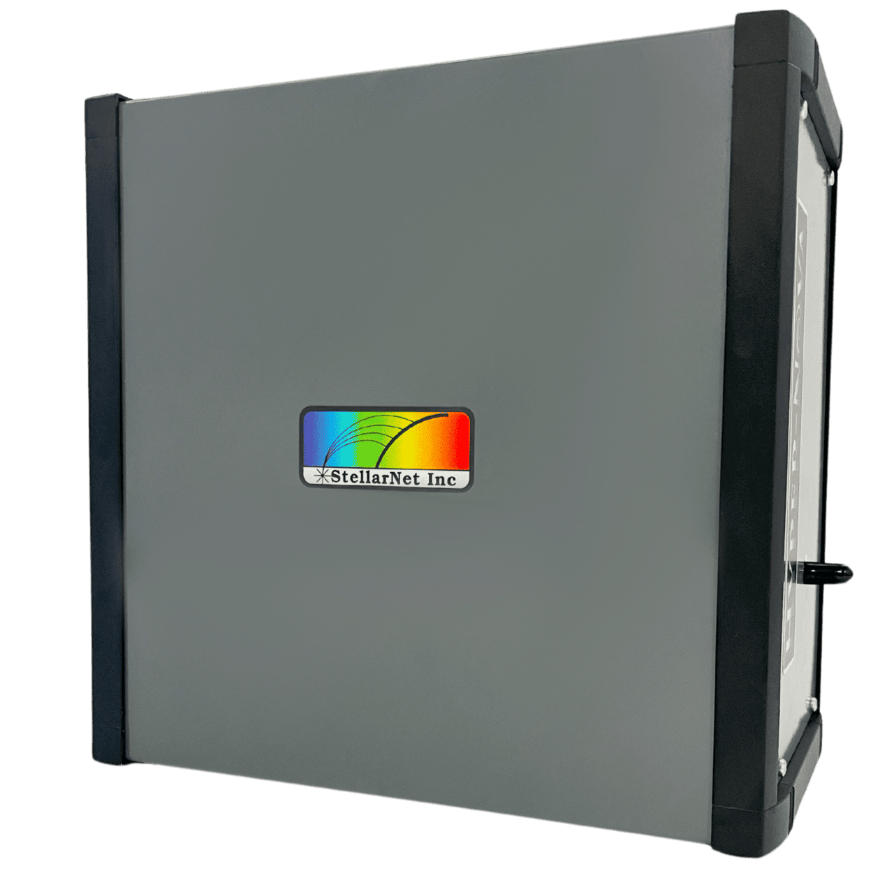
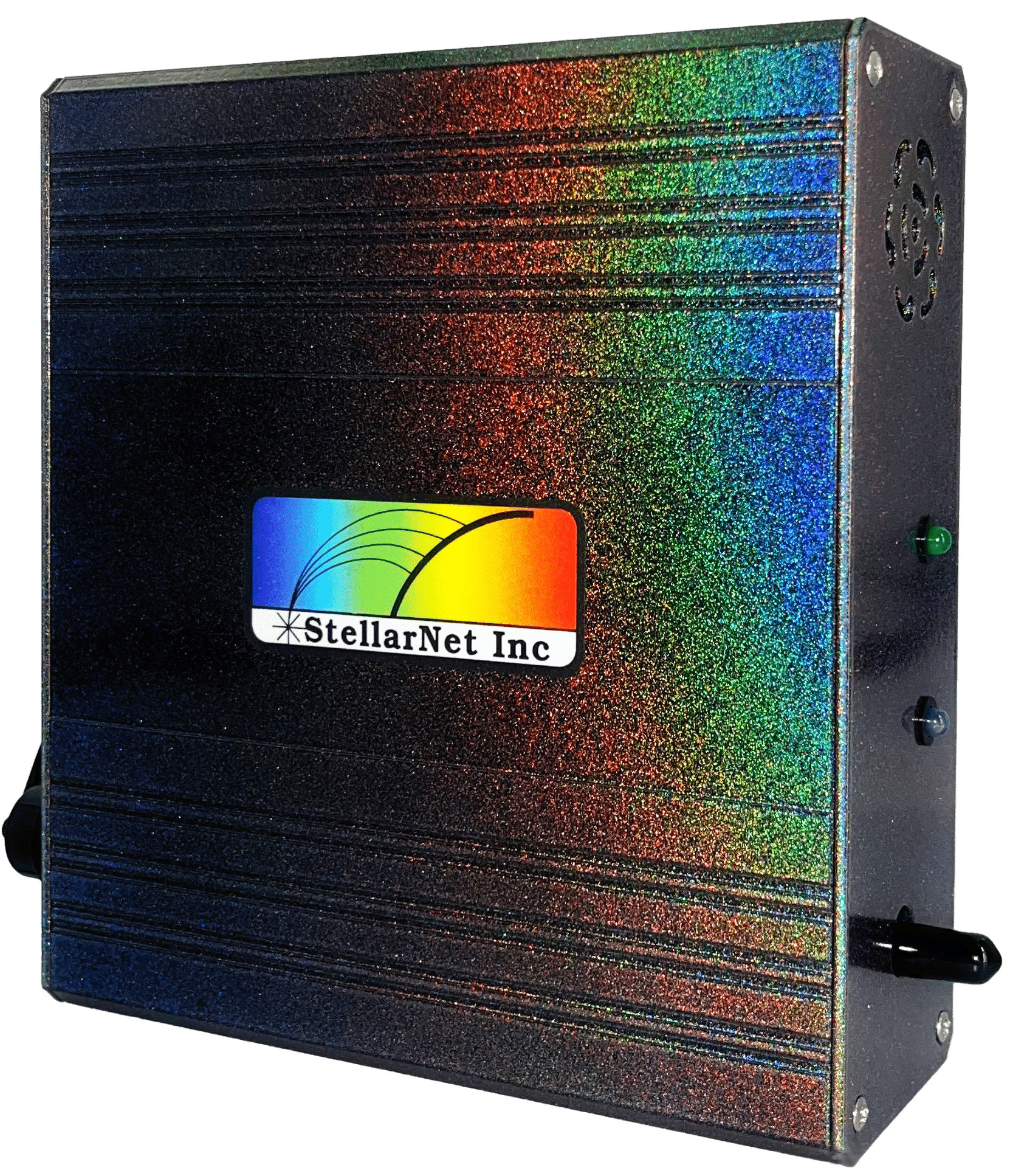
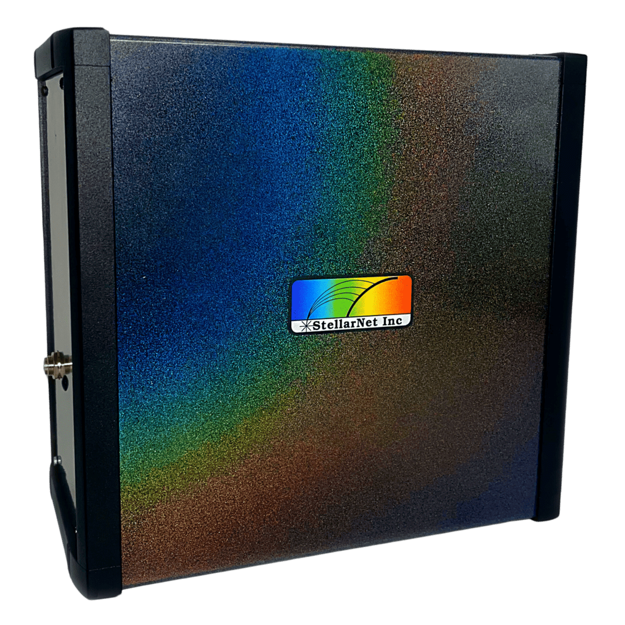
Mercury-OCT Engine: A Game-Changer in Imaging
The Mercury-OCT Engine by StellarNet offers unprecedented performance and affordability for OCT applications. Key features include:
- Fast Scanning Speed: Rapid data acquisition for real-time imaging, critical for dynamic processes.
- Broad Spectral Range: Enhanced imaging depth and contrast for various tissue types.
- Affordability: StellarNet is your low cost leader in spectroscopy solutions and brings the new Mercury-OCT Engine to market with leading price points.
- Multiple Mercury Configurations: Mercury-OCT-500 and Mercury-OCT 800 offer multiple wavelength ranges and imaging depths. Contact us for more information.
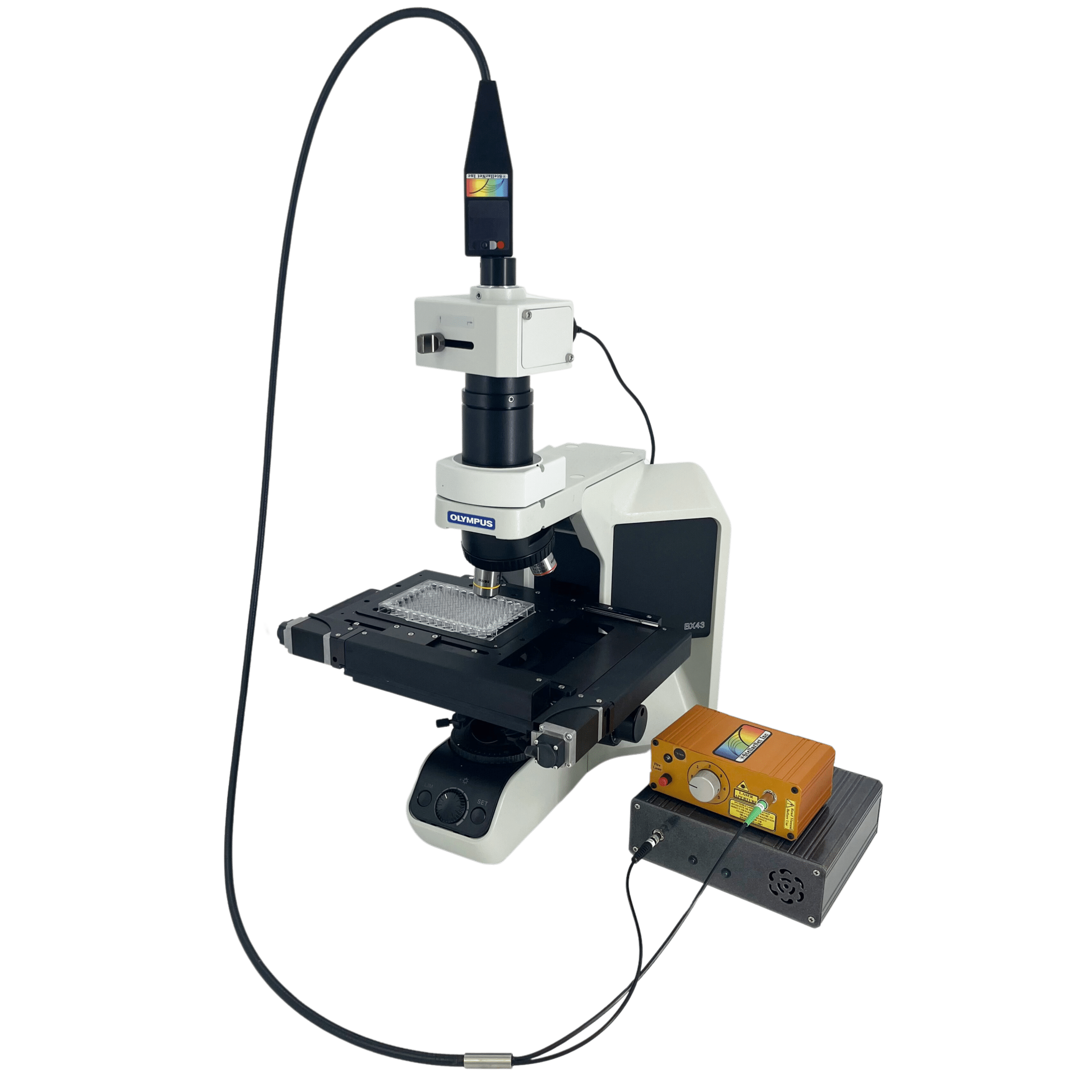
Complete SD (Spectral Domain) OCT System Hardware Components
To build a complete OCT system, the Mercury-OCT Engine integrates with several key hardware components:
- Fiber coupler: 50:50
- Light Source: A broadband light source is essential for illuminating the sample and obtaining detailed images.
- Scanner: A precision scanner moves the light beam across the sample, collecting comprehensive data.
- Interferometer: Splits the light into reference and sample arms, enabling the measurement of reflected light.
Software and Imaging Capabilities
Our new StellarPro software accompanies the Mercury-OCT Engine and collects spectral data with similar control and function as our standard spectrometers. We provide an SDK that allows users to further develop the collected data to generate high-resolution, cross-sectional images. These images can be rendered in both two-dimensional and three-dimensional formats, providing detailed insights into tissue morphology and pathology. Researchers and clinicians can use this data to analyze structural features, monitor disease progression, and guide therapeutic interventions.
For more information about the Mercury-OCT, please contact us here: